A bright cell stands out against a dark backdrop, resembling a flipbook where images blend seamlessly to depict the movement of an immune cell known as a phagocyte. This cell approaches a deceased cell, identifying it as a potential threat and engulfing it in a process referred to as phagocytosis.
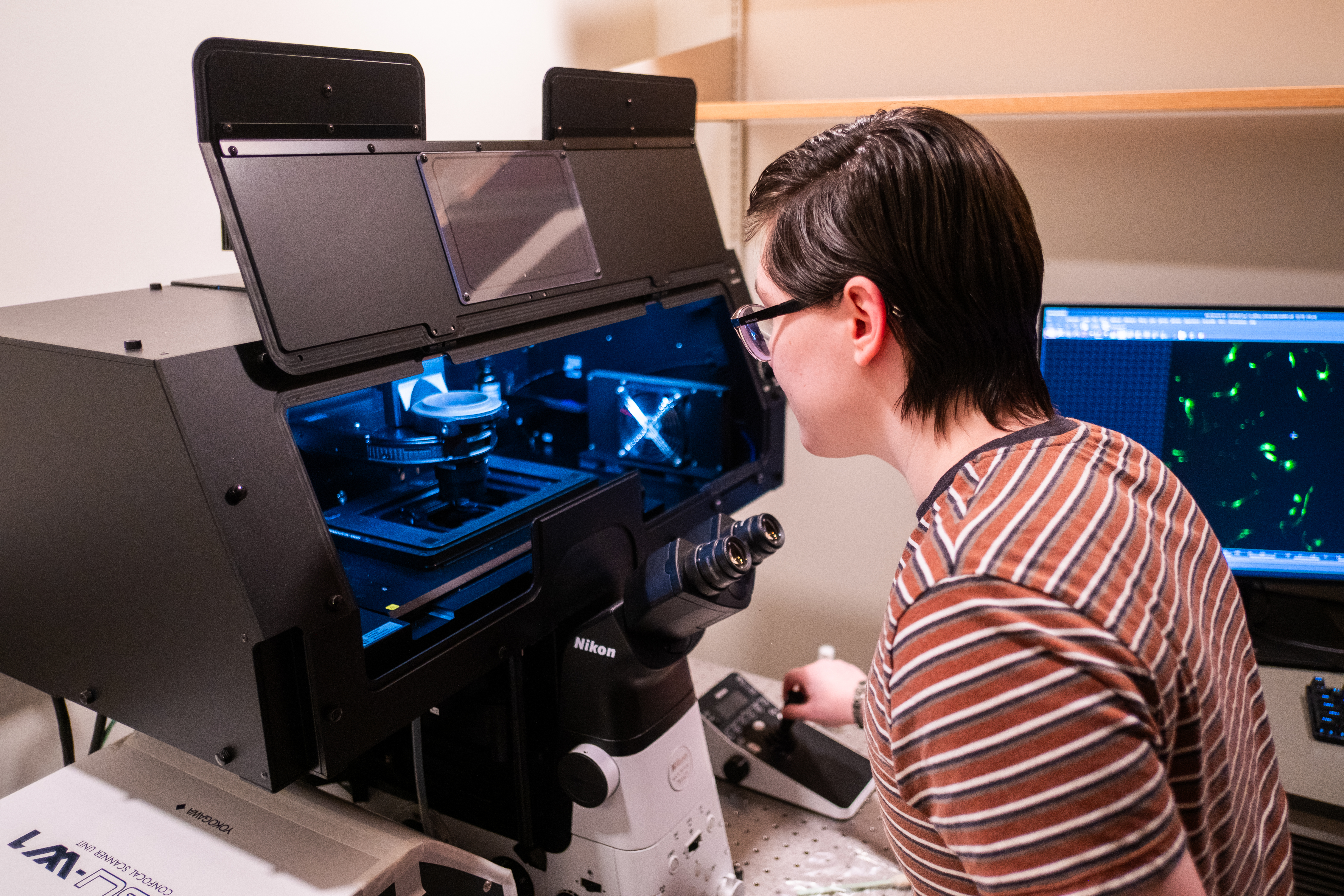
The process of phagocytosis typically occurs rapidly, often within minutes. The new spinning disk confocal microscope in the Park Science Building now enables researchers to observe this process moment by moment.
Assistant Professor of Biology Adam Williamson explains, “What sets this microscope apart is its capability to swiftly capture three-dimensional snapshots of active phagocytes with minimal harm to the cells under study, providing valuable insights into our research.”
Apart from its high-resolution imaging and real-time functionality, the confocal microscope features a controlled environment chamber for humidity and temperature. This feature allows scientists to work with live cells, a task previously unattainable with an electron microscope. The microscope is utilized by Williamson, Dean of Graduate Studies and Physics Professor May Cheng, Assistant Professor of Physics Asja Radja, and their respective students.
For Radja, the availability of the microscope was instrumental in her decision to join Bryn Mawr faculty in 2022. Her research focuses on biological patterns and shapes, ranging from macroscopic to microscopic levels, and the underlying physics governing their formation.
Radja’s study involves gorgonians, soft corals known for their resistance to bleaching compared to hard corals. These corals harbor polyps, individual organisms that Radja is examining alongside Villanova master’s student Michael Drummond.
Drummond expresses his amazement at the opportunity the microscope provides to observe algae living on the polyps in unprecedented detail. During his initial microscope usage, Drummond made a novel discovery of a unique algae pattern, a finding that excited Radja as it had not been documented before.
Before acquiring the confocal microscope, Williamson had to travel to the University of Delaware to access a similar tool. The inconvenience prompted the acquisition of the microscope, which Williamson describes as indispensable to their research endeavors.
Williamson collaborates with Kyle Bledsoe ‘24, a biochemistry and molecular biology major, on a project funded by an NIH grant. The project delves into how genes mutated in Batten Disease, a group of lysosomal storage disorders, safeguard the nervous system.
As they prepare human retinal cells for study using CRISPR gene editing technology, Williamson and Bledsoe aim to understand the protective mechanisms of the mutated genes in Batten Disease. Their ultimate goal is to determine if varying types of the disease necessitate distinct treatments based on cellular responses.
Despite the extensive preparations required before microscopy analysis, Bledsoe acknowledges the machine’s intricacy and versatility in exploring different light wavelengths. As Bledsoe’s research term nears its end, Williamson anticipates passing the baton to future students, emphasizing the thrill of witnessing groundbreaking scientific discoveries unfold before their eyes.
\ Get the latest news /