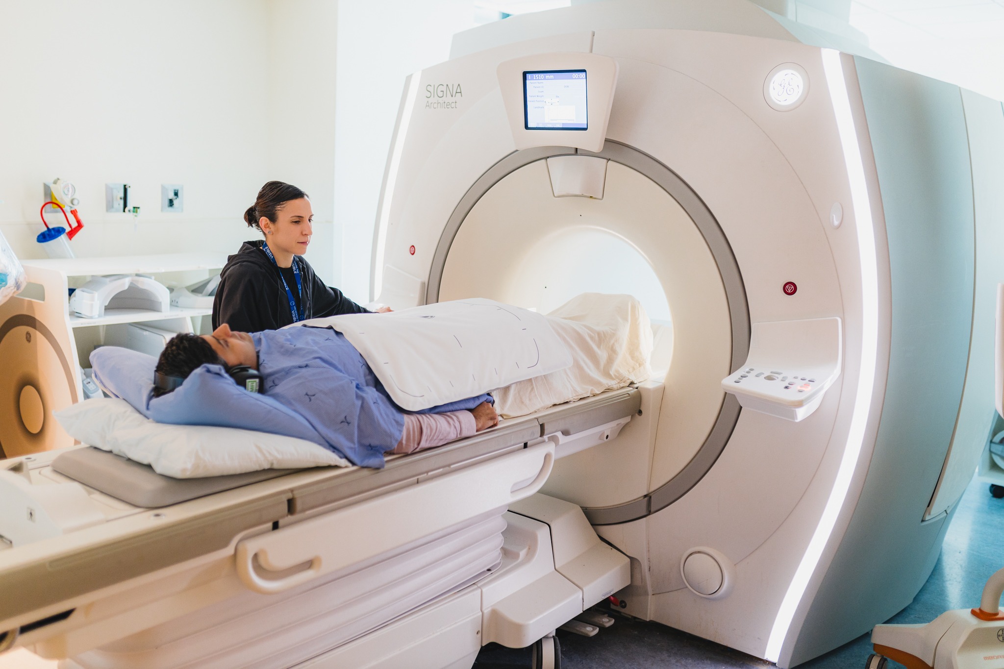
Senior MRI technologist Liz Teixeira and radiologist Dr. Ameya Madhav Kulkarni, who is the patient in the accompanying photo, are demonstrating an MRI-ultrasound fusion biopsy procedure now accessible at HHS Juravinski Hospital and Cancer Centre. This innovative diagnostic imaging technology is significantly enhancing the identification of prostate cancer, ensuring patients receive the most suitable treatment.
Advanced Biopsy Technique at JHCC
Hamilton Health Sciences (HHS) Juravinski Hospital and Cancer Centre has introduced cutting-edge diagnostic imaging technology to improve the accurate detection of prostate cancer, including high-risk variants, enabling personalized treatment for patients.
“By utilizing this biopsy system, we can precisely target cancer samples that may pose a life-threatening risk.” — Dr. Scott Tsai, Radiologist at HHS
Treatment for aggressive prostate cancers may involve surgical procedures, radiation therapy, and/or chemotherapy, whereas low-risk cases may only necessitate regular monitoring. The new MRI-ultrasound fusion biopsy system at JHCC merges MRI with ultrasound to generate detailed 3-D prostate images during biopsies.
Understanding Prostate Cancer
Statistics from the Canadian Cancer Society reveal that one in eight men in Canada will receive a prostate cancer diagnosis in their lifetime, making it the most prevalent cancer among Canadian men.

The team comprises Jeanine Risk, the Manager of Diagnostic Imaging at JHCC and Nuclear Medicine; Dr. Lori Stewart, a Staff Radiologist and Body Imaging Lead at JHCC; Dr. Scott Tsai, Radiologist; and Stephen Zuccolo, Ultrasound Technologist.
The majority of prostate cancers exhibit slow growth and remain localized within the prostate gland, often posing no immediate threat. Dr. Ameya Madhav Kulkarni, an HHS radiologist, explains that individuals diagnosed with low-risk prostate cancer may never experience symptoms throughout their lifetime.
However, certain aggressive prostate cancer types can metastasize rapidly, presenting a significant danger. Early detection of prostate cancer while it is still confined to the prostate gland offers the best prognosis for successful treatment.
Elevating Biopsy Standards
In Canada and various global regions, a systematic biopsy is the gold standard for diagnosing prostate cancer. This procedure involves extracting 10 to 12 tissue samples from random prostate areas using a needle. Ultrasound guidance assists in determining the sampling locations during systematic biopsies. Ultrasound technology utilizes sound waves to produce detailed images crucial for disease diagnosis, including cancer.

Dr. Ameya Madhav Kulkarni and Senior MRI Technologist Liz Teixeira
Tissue samples collected during biopsies are sent for testing to identify the cancer type. However, due to the random sampling nature of systematic biopsies, there is a risk of missing cancerous areas.
MRI technology utilizes magnetic fields and radio waves to produce cross-sectional images that can detect aggressive prostate cancer types, highlight cancerous areas overlooked by other tests, and ascertain the size, location, and potential spread of cancer.
The integration of MRI and ultrasound technologies generates synchronized 3-D prostate images, enabling radiologists to pinpoint specific biopsy sites within the prostate, thereby enhancing the detection of clinically significant cancers.
Dr. Scott Tsai, an HHS radiologist, emphasizes the advantage of MRI in detecting clinically relevant prostate cancers. He states, “By utilizing this biopsy system, we can precisely target cancer samples that may pose a life-threatening risk.”
HHS introduced this technology in November, providing comprehensive training to team members on the new system. HHS and St. Joseph Healthcare Hamilton are the exclusive hospitals in the region offering MRI-ultrasound fusion biopsies.
\ Get the latest news /